Southwest Diagnostic Imaging Center Uses Artificial Intelligence to Optimize Breast Tomosynthesis Interpretations


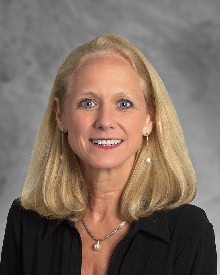
Dr. Katherine Hall, MD
Radiologist and Co-Director of Mammography, East Division, at Southwest Diagnostic Imaging Center
Challenge:
Responsible for reading nearly 12,000 mammograms each year.
Implementation of digital breast tomosynthesis (3D mammography solution).
Getting radiology team buy-in on a new solution leveraging artificial intelligence and deep learning.
Solution:
ProFound AI® for Digital Breast Tomosynthesis
Results:
Made the complete switch from 2D to 3D protocol.
Reduced reading time of up to 50%.
The Story of Southwest Diagnostic Imaging
Located in a spacious 25,000 square foot facility on the Presbyterian Hospital of Dallas campus, Southwest Diagnostic Imaging Center is one of the largest freestanding imaging centers in the Dallas-Fort Worth area. Since 1985, the facility has provided thousands of patients from Texas and across the nation with the highest standard of service and a patient-centered approach to care.
ProFound AI® is such a wonderful addition. When you’re confronted with a lot of breast tomosynthesis exams daily to read, you don’t want to get fatigued. Now, depending on the size of the breast, I am able to cut down my reading time anywhere from 25-50%. – Dr. Katherine Hall, Southwest Diagnostic Imaging Center
Leading the Way
While the adoption of breast tomosynthesis is expanding, so is the innovative technology which supports these solutions. Southwest Diagnostic Imaging Center has been paving the way, equipping its radiologists with the most advanced imaging solutions for more than 30 years, and has now added artificial intelligence solution ProFound AI to their women’s breast health program.
To maintain competitive advantage, transitioning from 2D to 3D mammography, or digital breast tomosynthesis (DBT), was the imaging center’s next step. However, with nearly 12,000 mammograms conducted each year, finding a cutting-edge solution that ensured both accuracy and efficiency was crucial.
“Previously running a facility that used DBT for many years, I knew first-hand the incredible benefits and challenges radiologists face with the technology,” said Dr. Hall, radiologist and co-director of mammography, east division, at Southwest Diagnostic Imaging Center. “While I was eager to make the switch from 2D to DBT to better serve our patients, I also expected some internal resistance, as going from reading just a few images in 2D to reading hundreds of images per patient with DBT can be both tiresome and time-intensive for radiologists.”
Conducting the Search
In May 2016 Southwest Diagnostic Imaging Center conducted an extensive review of all existing tomosynthesis technologies on the market, ultimately deciding to implement GE Healthcare’s DBT solution. In doing so, the facility was also able to take advantage of iCAD’s ProFound AI.
Specifically, ProFound AI reduces the need to scroll through hundreds of images. Rather, it utilizes deep learning to pick up the highest densities and areas of distortion on a mammogram, extracts them and blends them onto a synthetic 2D image.
“Instead of looking at 80-90 planes or more per projection for those patients with large breasts, ProFound AI leads me directly to the images that are most critical to review,” said Dr. Hall. “I am then able to click on that area in the preview and the solution takes me directly to the location needing further evaluation. In fact, any suspicious masses or distortions on the image are so clear that they literally pop out at you.”
Reducing Read Time, Improving Workflow
After four months, Southwest Diagnostic Imaging Center’s radiologists are reaping the benefits of ProFound AI. In fact, not only did ProFound AI help Dr. Hall move to an all 3D protocol, she’s also cut down her reading time anywhere from 25-50 percent. “We want to ensure quality care for our patients and with the adoption I feel confident to utilize a 3D only protocol. This is a benefit to our patients as it is a reduction in radiation dose exposure.”
“As radiologists, when we’re amassed with information and are in a state of image overload, we all need help seeing,” said Dr. Hall. “ProFound AI has been a game changer. It helps me collimate and focus on what I really need to look at and evaluate, which is what I rely on to provide my patients with the best possible care.”
Disclaimer: This case study represents the experience and position of the clinician and doesn’t represent the opinions of iCAD, Inc. Clinicians referenced in the study are responsible for the accuracy of provided data. Refer to the device user manual for detailed product information. To request a copy of the device user manual, please contact iCAD, Inc.
