

Science
The Science is ProFound.
Backed by science, clinical evidence, and proven patient outcomes.
Why imaging centers partner with iCAD.
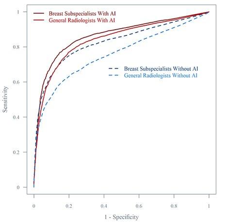
Radiologists using iCAD’s best-in-class artificial intelligence-powered solutions in our ProFound Breast Health Suite are achieving remarkable outcomes:
Cancer Detection
The world’s first and most reliable deep-learning AI mammography detection software, ProFound Detection empowers radiologists to advance their diagnostic accuracy and performance.4
The ProFound AI algorithm rapidly and accurately analyzes each individual image or slice and identifies potentially malignant lesions.
Summary:
- Trained on over 20 million images
- Data gathered from over 130 sites from around the globe
- Analyzes each slice for masses, distortion, calcifications & asymmetry
- Localizes, segments & classifies lesions
- Provides Lesion Score & Case Score for each lesion & exam
How does it work?
ProFound Detection’s deep-learning algorithm uses advanced neural network image processing and pattern recognition to analyze mammography images for potential areas of concern. Suspicious appearing structures such as densities (masses, architectural distortions, and asymmetries) and calcification clusters are identified via a contoured line.
Trained with one of the largest available 3D image datasets of over 20 million images, ProFound AI provides radiologists with crucial information, such as Case Scores, which assist in prioritizing caseload and clinical decision-making.
ProFound Detection V4 is FDA-cleared to offer superior performance and sensitivity to support radiologists in the fight against breast cancer with an unrivaled accuracy of detection.
ProFound AI® V3 is FDA-cleared, CE Marked, and Health Canada Licensed. Outside the United States, it’s a clinically proven solution designed to amplify radiologists’ diagnostic accuracy and performance reading 2D mammography and digital breast tomosynthesis (DBT).
The ProFound Scorecard provides an easy-to-read report including:
Lesion Score
Each identified region is assigned a localized score, indicating its match to a similar malignant lesion within ProFound’s AI data set
Case Score
Each case’s overall score indicates the algorithm’s confidence level for the case matching a malignant case within ProFound’s AI data set and used to prioritize reading worklist
Designed to assist with clinical decision making
Can be flexibly integrated into MIS/RIS software
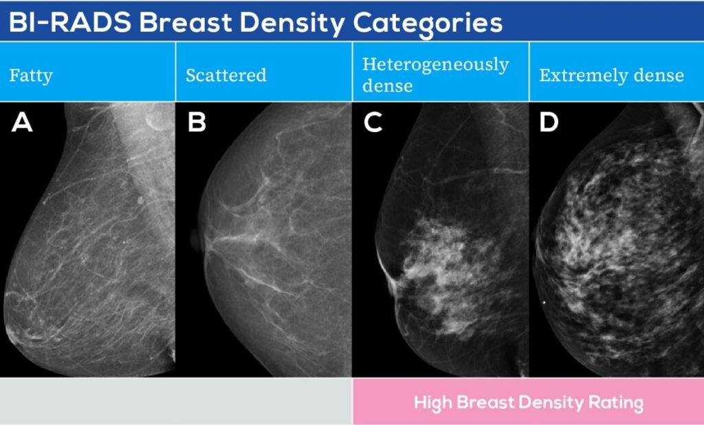
Density
iCAD’s Density Assessment software simplifies density assessment, reporting, and stratification with its accurate and reliable automated density categorization.
Summary:
- Objective, reliable, and consistent breast density assessments
- Highest matching accuracy for dense and non-dense assessment on the market11
- Seamless integration with multiple PACS, reading workstations, and RIS/MIS
How does it work?
Our Density Assessment solution identifies anatomy, segments the breast, then measures adipose and fibroglandular tissue and its dispersion to determine the density category in alignment to BI-RADS® lexicon.
It’s the world’s first multi-vendor automated breast density assessment algorithm powered by deep learning for 2D and 3D mammography systems, using synthesized 2D images.11
Integrate Seamlessly: Density Assessment results can be integrated into common risk models that incorporate density and automated mammography reports through leading MIS/RIS software products.
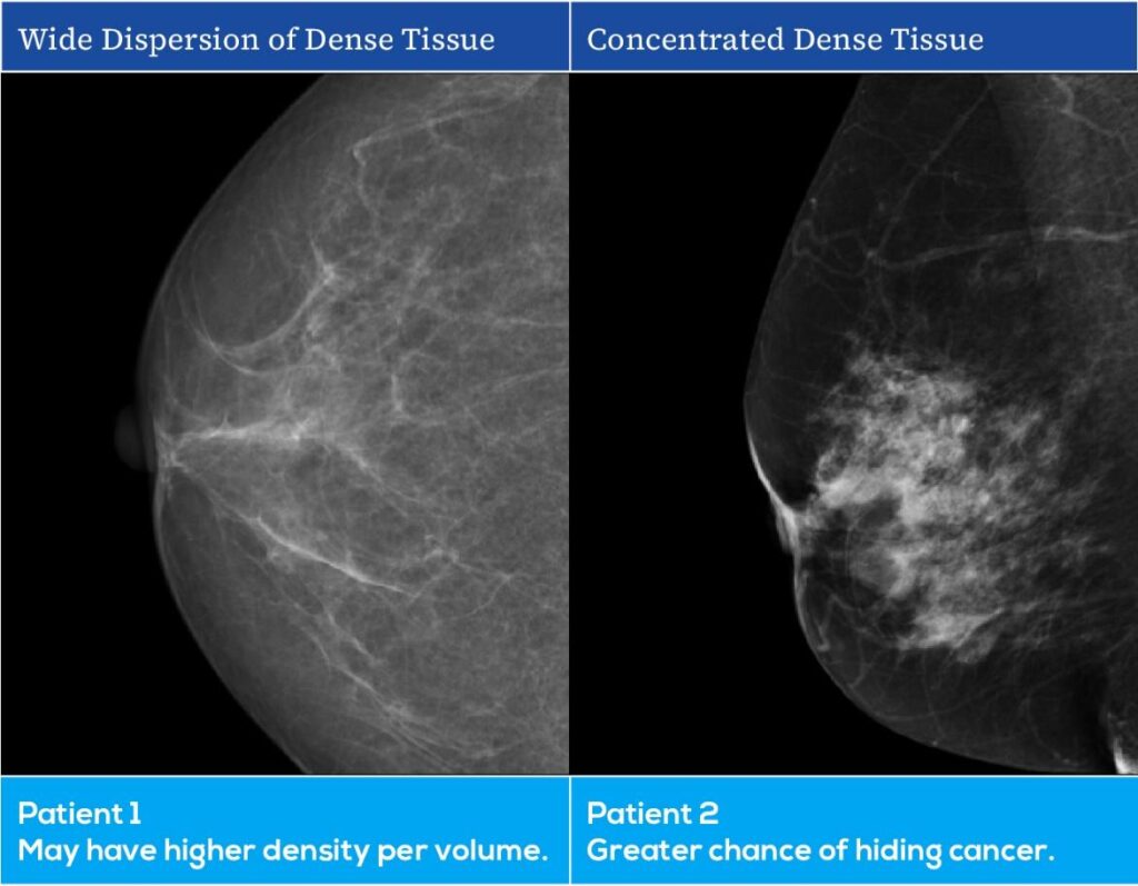
The dispersion or localized concentration of density affects how difficult it is to detect a lesion in the same rated density category.
Risk
ProFound AI® Risk is an accurate, easy to use breast cancer risk solution that enables clinicians to truly personalize screening by further stratifying breast cancer risk, resulting in personalized screening plans to identify cancer earlier.
ProFound AI Risk is CE Marked and Health Canada Licensed, is not FDA Cleared, and is only available in the US for investigational use.
Why do we need an updated way to evaluate risk?
- About 85% of breast cancers occur in women who have no family history of breast cancer.12
- Women with very dense breasts are 4-5 times more likely to get breast cancer than women with fatty breasts.13,14
Summary:
- World’s first image-informed risk model that identifies women at high risk of developing cancer before or at their next screening
- 2.4x more accurate than traditional lifestyle models, such as Tyrer-Cuzick1,2
- Complements traditional risk models and empowers clinicians to personalize screening regimens and find cancers earlier
- Offers similar performance in women with dense and non-dense breasts11
How does it work?
Applied to either a standard Full Field Digital Mammogram (FFDM) or combined Digital Breast Tomosynthesis exam (DBT+2D), the ProFound Risk solution leverages ProFound AI Detection breast complexity findings, ProFound Density, and age to calculate a woman’s short-term, absolute risk of breast cancer. ProFound Risk results, viewed within a hanging protocol, include the 1- or 2- year breast cancer risk score and breast cancer risk category (Low, General, Moderate and High), rooted with either NICE or USPSTF thresholds.
The easy to integrate ProFound Risk solution provides superior accuracy and enables clinicians to personalize patient screening to find breast cancer early.
Learn more about us.
1. Mikael Eriksson et al. A risk model for digital breast tomosynthesis to predict breast cancer and guide clinical care. Sci. Transl. Med. 14, eabn3971 (2022). DOI: 10.1126/scitranslmed.abn3971. 2. Eriksson M, Czene K, Strand F, Zackrisson S, Lindholm P, Lång K, Förnvik D, Sartor H, Mavaddat N, Easton D, Hall P. Identification of Women at High Risk of Breast Cancer Who Need Supplemental Screening. Radiology. 2020 Nov;297(2):327-333. doi: 10.1148/radiol.2020201620. Epub 2020 Sep 8. PMID: 32897160. 4. Conant EF, Toledano AY, Periaswamy S, Fotin SV, Go J, Boatsman JE, Hoffmeister JW. Improving Accuracy and Efficiency with Concurrent Use of Artificial Intelligence for Digital Breast Tomosynthesis. Radiol Artif Intell. 2019 Jul 31;1(4):e180096. doi: 10.1148/ryai.2019180096. PMID: 32076660; PMCID: PMC6677281. 13. Boyd NF, Guo H, Martin LJ, Sun L, Stone J, Fishell E, Jong RA, Hislop G, Chiarelli A, Minkin S, Yaffe MJ. Mammographic density and the risk and detection of breast cancer. N Engl J Med. 2007 Jan 18;356(3):227-36. doi: 10.1056/NEJMoa062790. PMID: 17229950. 14. Yaghjyan L, Colditz GA, Collins LC, Schnitt SJ, Rosner B, Vachon C, Tamimi RM. Mammographic breast density and subsequent risk of breast cancer in postmenopausal women according to tumor characteristics. J Natl Cancer Inst. 2011 Aug 3;103(15):1179-89. doi: 10.1093/jnci/djr225. Epub 2011 Jul 27. PMID: 21795664; PMCID: PMC3149043. 16. Lauby-Secretan B, Scoccianti C, Loomis D, Benbrahim-Tallaa L, Bouvard V, Bianchini F, Straif K; International Agency for Research on Cancer Handbook Working Group. Breast-cancer screening–viewpoint of the IARC Working Group. N Engl J Med. 2015 Jun 11;372(24):2353-8. doi: 10.1056/NEJMsr1504363. Epub 2015 Jun 3. PMID: 26039523.
iCAD data on file. Standalone performance varies by vendor.